How to cultivate oocytes with the Graphical representation tab?
From MedITEX - Wiki
| (12 intermediate revisions by 2 users not shown) | |||
| Line 1: | Line 1: | ||
| − | <p>The graphical | + | <p>The <strong>graphical representation</strong> allows the documentation of individual<strong> oocytes/embryos</strong>, its development status and oocyte-specific processes on all culture days.</p> |
| − | <table border="0"> | + | <table style="margin-left: auto; margin-right: auto;" border="0"> |
<tbody> | <tbody> | ||
<tr> | <tr> | ||
| Line 6: | Line 6: | ||
<td> | <td> | ||
<ul> | <ul> | ||
| − | <li> | + | <li><strong>MedITEX IVF</strong> adds oocytes/embryos obtained by <strong>follicle aspiration</strong> or <strong>thawing</strong> procedures.</li> |
| − | + | <li>You can document the development up to 10 days.</li> | |
| − | + | ||
| − | </li> | + | |
</ul> | </ul> | ||
</td> | </td> | ||
| Line 15: | Line 13: | ||
</tbody> | </tbody> | ||
</table> | </table> | ||
| − | <p>Within | + | <p> </p> |
| − | <table border="0"> | + | <p>Within this view, you have three options to edit the oocytes/embryos:</p> |
| + | <table style="margin-left: auto; margin-right: auto;" border="0" width="319" height="106"> | ||
<tbody> | <tbody> | ||
<tr> | <tr> | ||
| − | <td | + | <td colspan="2"><ol> |
| − | < | + | <li>Edit one or more fields with the <a href="/index.php?title=Cycle_Culture#1._Right-panel."><strong>right-panel</strong></a>.<strong> <br /></strong></li> |
| − | <li><strong>Double-click</strong> to enter development stages, scores, notes, photos, assisted hatching and polar body diagnostics. </li> | + | <li><a href="/index.php?title=Cycle_Culture#2._Double-click"><strong>Double-click</strong></a> to enter development <strong>stages</strong>, <strong>scores</strong>, <strong>notes</strong>, <strong>photos</strong>,<strong> assisted hatching</strong> and <strong>polar body diagnostics</strong>. </li> |
| − | <li><strong>Right-click</strong> to enter treatments, stages and modifications.</li> | + | <li><a href="/index.php?title=Cycle_Culture#3._Right-click"><strong>Right-click</strong></a> to enter <strong>treatments</strong>, <strong>stages </strong>and <strong>modifications</strong>.</li> |
</ol></td> | </ol></td> | ||
| + | </tr> | ||
| + | <tr> | ||
| + | <td><img src="/images/o0.png" alt="" /></td> | ||
| + | <td><strong>Oocytes/embryos</strong> added to the <strong>Graphical representation</strong> view, will initially display the following icon.<br /></td> | ||
</tr> | </tr> | ||
</tbody> | </tbody> | ||
</table> | </table> | ||
<p> </p> | <p> </p> | ||
| − | <h3> | + | <h3>Edit one or more fields with the vertical grid</h3> |
| − | <p> | + | <p>This is the quickest way of entering data. You can see all existing data of the <strong>selected oocyte/embryo</strong> and editing this information is simply performed by changing the related fields.<br />To edit the information for oocytes/embryos, select a cell from the <strong>Graphical representation </strong>view and add the relevant data to the <strong>Right-panel</strong>.</p> |
| − | <table border="0"> | + | <p>You can also edit multiple oocytes/embryos at the same time. Simply select several of them holding the <strong>CTRL</strong> or <strong>Shift </strong>keys (they must be in the same culture day). Keep in mind that the information added will be the same for all selected oocytes/embryos.</p> |
| + | <table style="margin-left: auto; margin-right: auto;" border="0"> | ||
<tbody> | <tbody> | ||
<tr> | <tr> | ||
| − | <td> | + | <td><img src="/images/vg1.png" alt="" width="765" /></td> |
</tr> | </tr> | ||
</tbody> | </tbody> | ||
</table> | </table> | ||
| − | <p> | + | <p> </p> |
| − | <table border="0"> | + | <p>Read more about the possibilities to <strong>select several fields of one day</strong> in the next section <a href="/index.php?title=Cycle_Culture#Select_several_oocytes_.2F_embryos"><strong>Select several oocytes/embryos</strong></a>.</p> |
| + | <p> </p> | ||
| + | <h3>Double-click on the oocyte<strong>/embryo</strong></h3> | ||
| + | <p>A new window will open in which you can enter required data:</p> | ||
| + | <table style="margin-left: auto; margin-right: auto;" border="0"> | ||
<tbody> | <tbody> | ||
<tr> | <tr> | ||
| − | <td><img src="/images/ | + | <td><img src="/images/graph10.png" alt="" width="692" height="716" /><br /></td> |
| − | + | ||
| − | + | ||
| − | </td> | + | |
</tr> | </tr> | ||
</tbody> | </tbody> | ||
</table> | </table> | ||
| − | <p> | + | <p> </p> |
| − | <p> | + | <p>The <strong>Add file</strong> functionality lets you insert as many pictures as you need. Additionally, once an image is added, you can magnify it by <strong>double-clicking </strong>it.</p> |
| − | <p> | + | <p>The tabs <strong>Assisted hatching</strong>, <strong>Polar body diagnosis </strong>and <strong>PGD</strong> have to be selected if those procedures were conducted. In the tabs, check the box for the corresponding procedures and enter remaining information.</p> |
| − | <table border="0"> | + | <table style="margin-left: auto; margin-right: auto;" border="0"> |
<tbody> | <tbody> | ||
<tr> | <tr> | ||
| Line 59: | Line 64: | ||
</tbody> | </tbody> | ||
</table> | </table> | ||
| − | <p> | + | <p> </p> |
| − | <p>Confirm your entries by clicking <strong> | + | <p>Confirm your entries by clicking <strong>OK</strong> and the appearance of the oocyte/embryo will be changed.</p> |
| − | <table border="0"> | + | <table style="margin-left: auto; margin-right: auto;" border="0"> |
<tbody> | <tbody> | ||
<tr> | <tr> | ||
| − | <td><img src="/images/graph5.png" alt=" | + | <td><img src="/images/graph5.png" alt="" /></td> |
</tr> | </tr> | ||
</tbody> | </tbody> | ||
</table> | </table> | ||
| − | |||
<p> </p> | <p> </p> | ||
| − | <h3>Right-click on the oocyte</h3> | + | <h3>Right-click on the oocyte/embryo</h3> |
| − | <p>After right-clicking the oocyte, a | + | <p>After <strong>right-clicking</strong> the oocyte/embryo, a menu will open in which you can select the changes on the development stage.</p> |
| − | <table border="0"> | + | <table style="margin-left: auto; margin-right: auto;" border="0"> |
<tbody> | <tbody> | ||
<tr> | <tr> | ||
<td><img src="/images/graph6.png" alt="" /></td> | <td><img src="/images/graph6.png" alt="" /></td> | ||
<td> | <td> | ||
| − | <p>All changes that are made here will be represented in the | + | <p><strong>All changes</strong> that are made here will be represented in the <strong>Table representation</strong> as well.</p> |
| + | <p>If you change the input method to the <strong>Table representation</strong> tab, the stages will not be recorded anymore.</p> | ||
</td> | </td> | ||
</tr> | </tr> | ||
</tbody> | </tbody> | ||
</table> | </table> | ||
| − | <p> | + | <p> </p> |
| − | <table border="0"> | + | <h3>States of oocytes</h3> |
| + | <table style="margin-left: auto; margin-right: auto;" border="0"> | ||
<tbody> | <tbody> | ||
<tr> | <tr> | ||
| − | <td> | + | <td><img src="/images/o0.png" alt="" width="55" height="38" /></td> |
| + | <td> | ||
| + | <p><strong>Unknown</strong>:</p> | ||
| + | <p>Oocytes that did not have their stage selected.</p> | ||
| + | </td> | ||
</tr> | </tr> | ||
| − | |||
| − | |||
| − | |||
| − | |||
| − | |||
| − | |||
| − | |||
<tr> | <tr> | ||
| − | <td><img src="/images/ | + | <td><img src="/images/o1.png" alt="" /></td> |
| + | <td> | ||
| + | <p><strong>Discarded</strong>:</p> | ||
| + | <p>Discarded oocytes are marked with a red cross.</p> | ||
| + | </td> | ||
</tr> | </tr> | ||
| − | |||
| − | |||
| − | |||
| − | |||
<tr> | <tr> | ||
| − | <td><img src="/images/ | + | <td><img src="/images/o2.png" alt="" /></td> |
<td> | <td> | ||
| − | <p> | + | <p><strong>Frozen</strong>:<strong><br /></strong></p> |
| + | <p style="text-align: justify;">Frozen oocytes are marked with a snowflake symbol.</p> | ||
</td> | </td> | ||
</tr> | </tr> | ||
| − | |||
| − | |||
| − | |||
| − | |||
| − | |||
| − | |||
<tr> | <tr> | ||
| − | <td><img src="/images/ | + | <td><img src="/images/o3.png" alt="" /></td> |
<td> | <td> | ||
| − | <p><strong> | + | <p><strong>Selected for transfer</strong>:</p> |
| − | <p> | + | <p style="text-align: justify;">Pro-nucleus stages that are selected for an embryo transfer are marked with a green circle. </p> |
</td> | </td> | ||
</tr> | </tr> | ||
<tr> | <tr> | ||
| − | <td><img src="/images/ | + | <td><img src="/images/o4.png" alt="" /></td> |
<td> | <td> | ||
| − | <p><strong> | + | <p><strong>Transferred</strong>:</p> |
| − | <p>Those that are to | + | <p>Those that are to be transferred are marked with the <strong>ET</strong> symbol.</p> |
</td> | </td> | ||
</tr> | </tr> | ||
<tr> | <tr> | ||
| − | <td><img src="/images/ | + | <td><img src="/images/o7.png" alt="" /></td> |
<td> | <td> | ||
| − | <p><strong> | + | <p><strong>In arrest</strong>:<strong> </strong></p> |
| − | <p> | + | <p>Oocytes that stopped developing.<strong><br /></strong></p> |
</td> | </td> | ||
</tr> | </tr> | ||
<tr> | <tr> | ||
| − | <td><img src="/images/ | + | <td><img src="/images/o5.png" alt="" /></td> |
<td> | <td> | ||
| − | <p><strong> | + | <p><strong>PBD</strong>:</p> |
| − | <p> | + | <p style="text-align: justify;">Oocytes that have been subjected to a polar body diagnosis.</p> |
</td> | </td> | ||
</tr> | </tr> | ||
<tr> | <tr> | ||
| − | <td><img src="/images/ | + | <td><img src="/images/o10.png" alt="" width="55" height="38" /></td> |
<td> | <td> | ||
| − | <p><strong> | + | <p><strong>PGD</strong>:</p> |
| − | <p>Oocytes that have been | + | <p>Oocytes that have been subjected to a pre-implantation genetic diagnosis.</p> |
</td> | </td> | ||
</tr> | </tr> | ||
<tr> | <tr> | ||
| − | <td><img src="/images/o6.png" alt=" | + | <td><img src="/images/o6.png" alt="" /></td> |
<td> | <td> | ||
| − | <p><strong>Assisted hatching | + | <p><strong>Assisted hatching</strong>:</p> |
| − | <p>Embryos | + | <p>Embryos subjected to assisted hatching.</p> |
</td> | </td> | ||
</tr> | </tr> | ||
<tr> | <tr> | ||
| − | <td><img | + | <td><img src="/images/o9.png" alt="" width="55" height="38" /></td> |
<td> | <td> | ||
| − | <p> | + | <p><strong>IVF</strong>:</p> |
| + | <p>Indicates the insemination method used.</p> | ||
| + | </td> | ||
| + | </tr> | ||
| + | <tr> | ||
| + | <td><img src="/images/o8.png" alt="" width="55" height="38" /></td> | ||
| + | <td> | ||
| + | <p><strong>ICSI</strong>:</p> | ||
| + | <p>Indicates the insemination method used.</p> | ||
</td> | </td> | ||
</tr> | </tr> | ||
| Line 165: | Line 171: | ||
</table> | </table> | ||
<p> </p> | <p> </p> | ||
| − | <p> | + | <h3>Treatment semen analysis</h3> |
| − | <table border="0"> | + | <p><strong>Double-click</strong> on the corresponding semen analysis. The window <strong>Treatment semen analysis</strong> will open.</p> |
| + | <table style="margin-left: auto; margin-right: auto;" border="0"> | ||
<tbody> | <tbody> | ||
<tr> | <tr> | ||
| − | <td> | + | <td>[[Image:Cul3.png|none|740px|thumb|left|link=http://wiki.meditex-software.com/images/Cul3.png| ]]</td> |
</tr> | </tr> | ||
</tbody> | </tbody> | ||
</table> | </table> | ||
<p> </p> | <p> </p> | ||
| − | + | <table style="margin-left: auto; margin-right: auto;" border="0"> | |
| − | <table border="0"> | + | |
<tbody> | <tbody> | ||
<tr> | <tr> | ||
| − | <td><img src="/images/ | + | <td><img src="/images/plus48.png" alt="" width="48" height="48" /></td> |
| + | <td><a href="/index.php?title=Treatments_and_treatment_semen_analysis">Click here</a> to have more information about the <strong>Treatment semen analysis</strong> window.</td> | ||
</tr> | </tr> | ||
</tbody> | </tbody> | ||
Latest revision as of 11:32, 15 April 2013
The graphical representation allows the documentation of individual oocytes/embryos, its development status and oocyte-specific processes on all culture days.
| <img src="/images/hint48.png" alt="" width="48" height="48" /> |
|
Within this view, you have three options to edit the oocytes/embryos:
|
|
| <img src="/images/o0.png" alt="" /> | Oocytes/embryos added to the Graphical representation view, will initially display the following icon. |
Contents |
Edit one or more fields with the vertical grid
This is the quickest way of entering data. You can see all existing data of the selected oocyte/embryo and editing this information is simply performed by changing the related fields.
To edit the information for oocytes/embryos, select a cell from the Graphical representation view and add the relevant data to the Right-panel.
You can also edit multiple oocytes/embryos at the same time. Simply select several of them holding the CTRL or Shift keys (they must be in the same culture day). Keep in mind that the information added will be the same for all selected oocytes/embryos.
| <img src="/images/vg1.png" alt="" width="765" /> |
Read more about the possibilities to select several fields of one day in the next section <a href="/index.php?title=Cycle_Culture#Select_several_oocytes_.2F_embryos">Select several oocytes/embryos</a>.
Double-click on the oocyte/embryo
A new window will open in which you can enter required data:
| <img src="/images/graph10.png" alt="" width="692" height="716" /> |
The Add file functionality lets you insert as many pictures as you need. Additionally, once an image is added, you can magnify it by double-clicking it.
The tabs Assisted hatching, Polar body diagnosis and PGD have to be selected if those procedures were conducted. In the tabs, check the box for the corresponding procedures and enter remaining information.
| <img src="/images/graph3.png" alt="" /> | <img src="/images/graph4.png" alt="" /> |
Confirm your entries by clicking OK and the appearance of the oocyte/embryo will be changed.
| <img src="/images/graph5.png" alt="" /> |
Right-click on the oocyte/embryo
After right-clicking the oocyte/embryo, a menu will open in which you can select the changes on the development stage.
| <img src="/images/graph6.png" alt="" /> |
All changes that are made here will be represented in the Table representation as well. If you change the input method to the Table representation tab, the stages will not be recorded anymore. |
States of oocytes
| <img src="/images/o0.png" alt="" width="55" height="38" /> |
Unknown: Oocytes that did not have their stage selected. |
| <img src="/images/o1.png" alt="" /> |
Discarded: Discarded oocytes are marked with a red cross. |
| <img src="/images/o2.png" alt="" /> |
Frozen: Frozen oocytes are marked with a snowflake symbol. |
| <img src="/images/o3.png" alt="" /> |
Selected for transfer: Pro-nucleus stages that are selected for an embryo transfer are marked with a green circle. |
| <img src="/images/o4.png" alt="" /> |
Transferred: Those that are to be transferred are marked with the ET symbol. |
| <img src="/images/o7.png" alt="" /> |
In arrest: Oocytes that stopped developing. |
| <img src="/images/o5.png" alt="" /> |
PBD: Oocytes that have been subjected to a polar body diagnosis. |
| <img src="/images/o10.png" alt="" width="55" height="38" /> |
PGD: Oocytes that have been subjected to a pre-implantation genetic diagnosis. |
| <img src="/images/o6.png" alt="" /> |
Assisted hatching: Embryos subjected to assisted hatching. |
| <img src="/images/o9.png" alt="" width="55" height="38" /> |
IVF: Indicates the insemination method used. |
| <img src="/images/o8.png" alt="" width="55" height="38" /> |
ICSI: Indicates the insemination method used. |
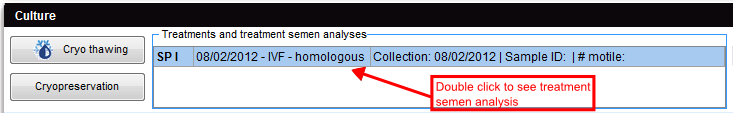
Treatment semen analysis
Double-click on the corresponding semen analysis. The window Treatment semen analysis will open.
 |
| <img src="/images/plus48.png" alt="" width="48" height="48" /> | <a href="/index.php?title=Treatments_and_treatment_semen_analysis">Click here</a> to have more information about the Treatment semen analysis window. |
| <a href="/index.php?title=MedITEX_IVF_how_to">Back to How to</a> | <a href="#top">Back to top</a> |