Come gestire la coltura con la Rappresentazione Grafica?
From MedITEX - Wiki
| Line 1: | Line 1: | ||
| − | <p> | + | <p><span>La </span><strong>Rappresentazione Grafica</strong><span> permette la documentazione di singoli </span><strong>ovociti/embrioni</strong><span>, del loro stadio di sviluppo e trattamenti specifice effettuati in qualsiasi giorno della coltura.</span></p> |
<table style="margin-left: auto; margin-right: auto;" border="0"> | <table style="margin-left: auto; margin-right: auto;" border="0"> | ||
<tbody> | <tbody> | ||
| Line 6: | Line 6: | ||
<td> | <td> | ||
<ul> | <ul> | ||
| − | <li><strong>MedITEX IVF</strong> | + | <li><strong>MedITEX IVF</strong> aggiungerà automaticamente le cellule aspirate durante il <strong>Pick Up</strong> o <strong>Scongelate</strong>.</li> |
| − | <li> | + | <li>Sarà possibile documentare un massimo di 10 giorni di coltura.</li> |
</ul> | </ul> | ||
</td> | </td> | ||
| Line 14: | Line 14: | ||
</table> | </table> | ||
<p> </p> | <p> </p> | ||
| − | <p> | + | <p>In questa sezione ci sono tre opzioni per inserire informazioni riguardanti le cellule:</p> |
<table style="margin-left: auto; margin-right: auto;" border="0" width="319" height="106"> | <table style="margin-left: auto; margin-right: auto;" border="0" width="319" height="106"> | ||
<tbody> | <tbody> | ||
<tr> | <tr> | ||
<td colspan="2"><ol> | <td colspan="2"><ol> | ||
| − | <li> | + | <li>Modificare uno o più campi dal <a href="/index.php?title=Coltura#1._Pannello_di_destra."><strong>pannello di destra.</strong></a><strong><br /></strong></li> |
| − | <li><a href="/index.php?title= | + | <li><a href="/index.php?title=Coltura#2._Doppio-click"><strong>Doppio-click</strong></a> per inserire informazioni sullo <strong>stadio, punteggio, note, foto e assisted hatching .</strong> </li> |
| − | <li><a href="/index.php?title= | + | <li>Cliccare con il <strong><a href="/index.php?title=Coltura#3._Click_tasto_destro">Tasto destro</a></strong> per inserire <strong>trattamenti</strong>, <strong>stadi ed eventuale transfer o crioconservazone.</strong></li> |
</ol></td> | </ol></td> | ||
</tr> | </tr> | ||
<tr> | <tr> | ||
<td><img src="/images/o0.png" alt="" /></td> | <td><img src="/images/o0.png" alt="" /></td> | ||
| − | <td><strong> | + | <td> |
| + | <p><strong>Ovociti/embrioni</strong> inseriti nella <strong>Rappresentazione Grafica</strong>, verranno visualizzati inizialmente con questa icona.</p> | ||
| + | </td> | ||
</tr> | </tr> | ||
</tbody> | </tbody> | ||
</table> | </table> | ||
<p> </p> | <p> </p> | ||
| − | <h3> | + | <h3>Modificare uno o più campi dalla griglia verticale</h3> |
| − | <p> | + | <p>Questa griglia è il metodo più veloce per inserire i dati. Sono elencati tutti i campi disponibili per le <strong>cellule selezionate </strong>e la modifica di tali informazioni è effettuata semplicemente modicicando il valore sei vari campi.<br />Per modificare le informazioni sarà quindi sufficiente selezionare una o più cellule (tenendo premuto <strong>CTRL</strong> o <strong>Maiusc</strong>) e agire sulla griglia.</p> |
| − | <p> | + | <p> </p> |
<table style="margin-left: auto; margin-right: auto;" border="0"> | <table style="margin-left: auto; margin-right: auto;" border="0"> | ||
<tbody> | <tbody> | ||
<tr> | <tr> | ||
| − | <td><img src="/images/ | + | <td><img src="/images/Coltura_21_IT.PNG" alt="" width="747" height="492" /></td> |
</tr> | </tr> | ||
</tbody> | </tbody> | ||
</table> | </table> | ||
<p> </p> | <p> </p> | ||
| − | <p> | + | <p>Per ulteriori informazioni su come <strong>selezionare più di una cellula alla volta appartenenti allo stesso giorno di coltura <a href="/index.php?title=Coltura#Selezionare_diversi_ovociti.2Fembrioni">Cliccare qui</a>.</strong></p> |
<p> </p> | <p> </p> | ||
| − | <h3> | + | <h3>Doppio-click su ovociti/embrioni</h3> |
| − | <p> | + | <p>La finestra seguente si aprirà:</p> |
<table style="margin-left: auto; margin-right: auto;" border="0"> | <table style="margin-left: auto; margin-right: auto;" border="0"> | ||
<tbody> | <tbody> | ||
<tr> | <tr> | ||
| − | <td><img src="/images/ | + | <td><img src="/images/Coltura_22_IT.PNG" alt="" width="764" height="759" /><br /></td> |
</tr> | </tr> | ||
</tbody> | </tbody> | ||
</table> | </table> | ||
<p> </p> | <p> </p> | ||
| − | <p> | + | <p>Premendo su <strong>Aggiungi file</strong> si ha la possibilità di inserire immagini relative alla cellula e successivamente ingrandirle facendo <strong>doppio-click.</strong></p> |
| − | <p> | + | <p>La tab <strong>Assisted hatching </strong>permette di inserire ulteriori informazioni riguardo questa procedura. Per attivare la sezione il check deve essere selezionato.</p> |
<table style="margin-left: auto; margin-right: auto;" border="0"> | <table style="margin-left: auto; margin-right: auto;" border="0"> | ||
<tbody> | <tbody> | ||
<tr> | <tr> | ||
<td><img src="/images/graph3.png" alt="" /></td> | <td><img src="/images/graph3.png" alt="" /></td> | ||
| − | |||
</tr> | </tr> | ||
</tbody> | </tbody> | ||
</table> | </table> | ||
<p> </p> | <p> </p> | ||
| − | <p> | + | <p>Per confermare premere <strong>OK</strong> e l'icona della cellula cambierà in conseguenza delle informazioni inserite.</p> |
<table style="margin-left: auto; margin-right: auto;" border="0"> | <table style="margin-left: auto; margin-right: auto;" border="0"> | ||
<tbody> | <tbody> | ||
| Line 74: | Line 75: | ||
</table> | </table> | ||
<p> </p> | <p> </p> | ||
| − | <h3> | + | <h3>Click con il tasto destro su ovociti/embrioni</h3> |
| − | <p> | + | <p>Cliccando su una cellula con il tasto destro il menù in figura apparirà:</p> |
<table style="margin-left: auto; margin-right: auto;" border="0"> | <table style="margin-left: auto; margin-right: auto;" border="0"> | ||
<tbody> | <tbody> | ||
| Line 81: | Line 82: | ||
<td><img src="/images/graph6.png" alt="" /></td> | <td><img src="/images/graph6.png" alt="" /></td> | ||
<td> | <td> | ||
| − | <p><strong> | + | <p><strong>Tutti i cambiamenti</strong><span> effettuati qui verranno automaticamente inseriti nella </span><strong>rappresentazione tabellare</strong><span>! </span></p> |
| − | <p> | + | <p>Se si riattiva la <strong>rappresentazione tabellare </strong>lo stadio delle cellule verrà perso.</p> |
</td> | </td> | ||
</tr> | </tr> | ||
| Line 88: | Line 89: | ||
</table> | </table> | ||
<p> </p> | <p> </p> | ||
| − | <h3> | + | <h3>Stati delle cellule</h3> |
<table style="margin-left: auto; margin-right: auto;" border="0"> | <table style="margin-left: auto; margin-right: auto;" border="0"> | ||
<tbody> | <tbody> | ||
| Line 94: | Line 95: | ||
<td><img src="/images/o0.png" alt="" width="55" height="38" /></td> | <td><img src="/images/o0.png" alt="" width="55" height="38" /></td> | ||
<td> | <td> | ||
| − | <p><strong> | + | <p><strong>Sconosciuta</strong>:</p> |
| − | <p> | + | <p>Cellule per le quali nessuno stadio è stato associato.</p> |
</td> | </td> | ||
</tr> | </tr> | ||
| Line 101: | Line 102: | ||
<td><img src="/images/o1.png" alt="" /></td> | <td><img src="/images/o1.png" alt="" /></td> | ||
<td> | <td> | ||
| − | <p><strong> | + | <p><strong>Scartata</strong>:</p> |
| − | <p> | + | <p>Cellule scartate sono contrassegnate da una x rossa.</p> |
</td> | </td> | ||
</tr> | </tr> | ||
| Line 108: | Line 109: | ||
<td><img src="/images/o2.png" alt="" /></td> | <td><img src="/images/o2.png" alt="" /></td> | ||
<td> | <td> | ||
| − | <p><strong> | + | <p><strong>Crioconservata</strong>:<strong><br /></strong></p> |
| − | <p | + | <p>Cellule crioconservate sono contrassegnate da un fiocco di neve.</p> |
| + | </td> | ||
| + | </tr> | ||
| + | <tr> | ||
| + | <td><img src="/images/thaw.PNG" alt="" width="55" height="38" /></td> | ||
| + | <td> | ||
| + | <p><strong>Scongelata:</strong></p> | ||
| + | <p>Cellule scongelate sono contrassegnate da un fiocco di neve sciolto.</p> | ||
</td> | </td> | ||
</tr> | </tr> | ||
| Line 115: | Line 123: | ||
<td><img src="/images/o3.png" alt="" /></td> | <td><img src="/images/o3.png" alt="" /></td> | ||
<td> | <td> | ||
| − | <p><strong> | + | <p><strong>Selezionata per transfer</strong>:</p> |
| − | <p | + | <p>2PN selezionabili per il transfer sono contrassegnati da un cerchio verde. </p> |
| + | </td> | ||
| + | </tr> | ||
| + | <tr> | ||
| + | <td><img src="/images/group.png" alt="" width="55" height="38" /></td> | ||
| + | <td> | ||
| + | <p><strong>Gruppi ovociti:</strong></p> | ||
| + | <p>Se sulla destra della cellula sono presenti delle linee colorate, la cellula fà parte di un gruppo di ovociti/embrioni.</p> | ||
</td> | </td> | ||
</tr> | </tr> | ||
| Line 122: | Line 137: | ||
<td><img src="/images/o4.png" alt="" /></td> | <td><img src="/images/o4.png" alt="" /></td> | ||
<td> | <td> | ||
| − | <p><strong> | + | <p><strong>Morfologia ideale per transfer</strong>:</p> |
| − | <p> | + | <p>Cellule, con morfologia ideale, da trasferire sono contrassegnate con il simbolo <strong>ET</strong> (verde).</p> |
| + | </td> | ||
| + | </tr> | ||
| + | <tr> | ||
| + | <td><img src="/images/et.PNG" alt="" width="55" height="38" /></td> | ||
| + | <td> | ||
| + | <p><strong>Morfologia non ideale per transfer</strong>:</p> | ||
| + | <p>Cellule, con morfologia non ideale, da trasferire sono contrassegnate con il simbolo <strong>ET</strong> (arancione).</p> | ||
</td> | </td> | ||
</tr> | </tr> | ||
| Line 129: | Line 151: | ||
<td><img src="/images/o7.png" alt="" /></td> | <td><img src="/images/o7.png" alt="" /></td> | ||
<td> | <td> | ||
| − | <p><strong>In | + | <p><strong>In arresto</strong>:<strong> </strong></p> |
| − | <p> | + | <p>Cellule che hanno smesso di svilupparsi.<strong><br /></strong></p> |
| + | </td> | ||
| + | </tr> | ||
| + | <tr> | ||
| + | <td><img src="/images/Donation_Donated.png" alt="" width="55" height="38" /></td> | ||
| + | <td> | ||
| + | <p><strong>Donazione - Donata:</strong></p> | ||
| + | <p>Cellule donate sono contrassegnate con una freccia verde a destra.</p> | ||
| + | </td> | ||
| + | </tr> | ||
| + | <tr> | ||
| + | <td><img src="/images/Donation_received.png" alt="" width="55" height="38" /></td> | ||
| + | <td> | ||
| + | <p><strong>Donazione - Ricevuta:</strong></p> | ||
| + | <p>Cellule ricevute sono contrassegnate con una freccia verde a sinistra.</p> | ||
</td> | </td> | ||
</tr> | </tr> | ||
| Line 137: | Line 173: | ||
<td> | <td> | ||
<p><strong>PBD</strong>:</p> | <p><strong>PBD</strong>:</p> | ||
| − | <p | + | <p>Cellule soggette a diagnosi del corpo polare.</p> |
</td> | </td> | ||
</tr> | </tr> | ||
<tr> | <tr> | ||
<td><img src="/images/o10.png" alt="" width="55" height="38" /></td> | <td><img src="/images/o10.png" alt="" width="55" height="38" /></td> | ||
| − | <td> | + | <td style="text-align: left;"> |
| − | <p><strong>PGD</strong>:</p> | + | <p><strong>PGD-PGS</strong>:</p> |
| − | <p> | + | <p>Cellule soggette a diagnosi/screening genetica/o pre-impianto.</p> |
</td> | </td> | ||
</tr> | </tr> | ||
<tr> | <tr> | ||
<td><img src="/images/o6.png" alt="" /></td> | <td><img src="/images/o6.png" alt="" /></td> | ||
| − | <td> | + | <td style="text-align: left;"> |
<p><strong>Assisted hatching</strong>:</p> | <p><strong>Assisted hatching</strong>:</p> | ||
| − | <p> | + | <p>Celllule sottoposte all' assisted hatching.</p> |
</td> | </td> | ||
</tr> | </tr> | ||
<tr> | <tr> | ||
<td><img src="/images/o9.png" alt="" width="55" height="38" /></td> | <td><img src="/images/o9.png" alt="" width="55" height="38" /></td> | ||
| − | <td> | + | <td style="text-align: left;"> |
<p><strong>IVF</strong>:</p> | <p><strong>IVF</strong>:</p> | ||
| − | <p> | + | <p>Metodo utilizzato per l'inseminazione.</p> |
</td> | </td> | ||
</tr> | </tr> | ||
<tr> | <tr> | ||
<td><img src="/images/o8.png" alt="" width="55" height="38" /></td> | <td><img src="/images/o8.png" alt="" width="55" height="38" /></td> | ||
| − | <td> | + | <td style="text-align: left;"> |
<p><strong>ICSI</strong>:</p> | <p><strong>ICSI</strong>:</p> | ||
| − | <p> | + | <p>Metodo utilizzato per l'inseminazione.</p> |
</td> | </td> | ||
</tr> | </tr> | ||
| Line 171: | Line 207: | ||
</table> | </table> | ||
<p> </p> | <p> </p> | ||
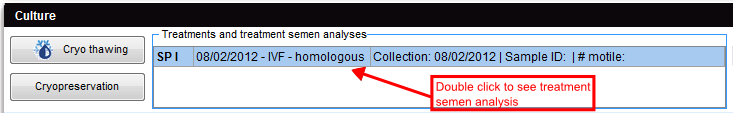
| − | <h3> | + | <h3>Trattamenti e Spermiogrammi associati </h3> |
| − | <p><strong> | + | <p>Facendo <strong>doppio click</strong> sulla riga del trattamento trattamento desiderato, la finestra di associazione dello spermiogramma si aprirà.</p> |
<table style="margin-left: auto; margin-right: auto;" border="0"> | <table style="margin-left: auto; margin-right: auto;" border="0"> | ||
<tbody> | <tbody> | ||
| Line 185: | Line 221: | ||
<tr> | <tr> | ||
<td><img src="/images/plus48.png" alt="" width="48" height="48" /></td> | <td><img src="/images/plus48.png" alt="" width="48" height="48" /></td> | ||
| − | <td><a href="/index.php?title= | + | <td><a href="/index.php?title=Trattamenti_e_spermiogrammi_associati">Cliccare qui</a> per maggiori informazioni sulla sezione dei Trattamenti e Spermiogrammi associati alla Coltura.</td> |
</tr> | </tr> | ||
</tbody> | </tbody> | ||
| Line 192: | Line 228: | ||
<tbody> | <tbody> | ||
<tr> | <tr> | ||
| − | <td style="text-align: right;"><a href="/index.php?title= | + | <td style="text-align: right;"><a href="/index.php?title=MedITEX_IVF_come_fare">Torna a Come fare</a></td> |
| − | <td style="text-align: right;"><a href="#top"> | + | <td style="text-align: right;"><a href="#top">Inizio pagina</a></td> |
</tr> | </tr> | ||
</tbody> | </tbody> | ||
</table> | </table> | ||
Revision as of 12:44, 2 May 2016
La Rappresentazione Grafica permette la documentazione di singoli ovociti/embrioni, del loro stadio di sviluppo e trattamenti specifice effettuati in qualsiasi giorno della coltura.
| <img src="/images/hint48.png" alt="" width="48" height="48" /> |
|
In questa sezione ci sono tre opzioni per inserire informazioni riguardanti le cellule:
|
|
| <img src="/images/o0.png" alt="" /> |
Ovociti/embrioni inseriti nella Rappresentazione Grafica, verranno visualizzati inizialmente con questa icona. |
Contents |
Modificare uno o più campi dalla griglia verticale
Questa griglia è il metodo più veloce per inserire i dati. Sono elencati tutti i campi disponibili per le cellule selezionate e la modifica di tali informazioni è effettuata semplicemente modicicando il valore sei vari campi.
Per modificare le informazioni sarà quindi sufficiente selezionare una o più cellule (tenendo premuto CTRL o Maiusc) e agire sulla griglia.
| <img src="/images/Coltura_21_IT.PNG" alt="" width="747" height="492" /> |
Per ulteriori informazioni su come selezionare più di una cellula alla volta appartenenti allo stesso giorno di coltura <a href="/index.php?title=Coltura#Selezionare_diversi_ovociti.2Fembrioni">Cliccare qui</a>.
Doppio-click su ovociti/embrioni
La finestra seguente si aprirà:
| <img src="/images/Coltura_22_IT.PNG" alt="" width="764" height="759" /> |
Premendo su Aggiungi file si ha la possibilità di inserire immagini relative alla cellula e successivamente ingrandirle facendo doppio-click.
La tab Assisted hatching permette di inserire ulteriori informazioni riguardo questa procedura. Per attivare la sezione il check deve essere selezionato.
| <img src="/images/graph3.png" alt="" /> |
Per confermare premere OK e l'icona della cellula cambierà in conseguenza delle informazioni inserite.
| <img src="/images/graph5.png" alt="" /> |
Click con il tasto destro su ovociti/embrioni
Cliccando su una cellula con il tasto destro il menù in figura apparirà:
| <img src="/images/graph6.png" alt="" /> |
Tutti i cambiamenti effettuati qui verranno automaticamente inseriti nella rappresentazione tabellare! Se si riattiva la rappresentazione tabellare lo stadio delle cellule verrà perso. |
Stati delle cellule
| <img src="/images/o0.png" alt="" width="55" height="38" /> |
Sconosciuta: Cellule per le quali nessuno stadio è stato associato. |
| <img src="/images/o1.png" alt="" /> |
Scartata: Cellule scartate sono contrassegnate da una x rossa. |
| <img src="/images/o2.png" alt="" /> |
Crioconservata: Cellule crioconservate sono contrassegnate da un fiocco di neve. |
| <img src="/images/thaw.PNG" alt="" width="55" height="38" /> |
Scongelata: Cellule scongelate sono contrassegnate da un fiocco di neve sciolto. |
| <img src="/images/o3.png" alt="" /> |
Selezionata per transfer: 2PN selezionabili per il transfer sono contrassegnati da un cerchio verde. |
| <img src="/images/group.png" alt="" width="55" height="38" /> |
Gruppi ovociti: Se sulla destra della cellula sono presenti delle linee colorate, la cellula fà parte di un gruppo di ovociti/embrioni. |
| <img src="/images/o4.png" alt="" /> |
Morfologia ideale per transfer: Cellule, con morfologia ideale, da trasferire sono contrassegnate con il simbolo ET (verde). |
| <img src="/images/et.PNG" alt="" width="55" height="38" /> |
Morfologia non ideale per transfer: Cellule, con morfologia non ideale, da trasferire sono contrassegnate con il simbolo ET (arancione). |
| <img src="/images/o7.png" alt="" /> |
In arresto: Cellule che hanno smesso di svilupparsi. |
| <img src="/images/Donation_Donated.png" alt="" width="55" height="38" /> |
Donazione - Donata: Cellule donate sono contrassegnate con una freccia verde a destra. |
| <img src="/images/Donation_received.png" alt="" width="55" height="38" /> |
Donazione - Ricevuta: Cellule ricevute sono contrassegnate con una freccia verde a sinistra. |
| <img src="/images/o5.png" alt="" /> |
PBD: Cellule soggette a diagnosi del corpo polare. |
| <img src="/images/o10.png" alt="" width="55" height="38" /> |
PGD-PGS: Cellule soggette a diagnosi/screening genetica/o pre-impianto. |
| <img src="/images/o6.png" alt="" /> |
Assisted hatching: Celllule sottoposte all' assisted hatching. |
| <img src="/images/o9.png" alt="" width="55" height="38" /> |
IVF: Metodo utilizzato per l'inseminazione. |
| <img src="/images/o8.png" alt="" width="55" height="38" /> |
ICSI: Metodo utilizzato per l'inseminazione. |
Trattamenti e Spermiogrammi associati
Facendo doppio click sulla riga del trattamento trattamento desiderato, la finestra di associazione dello spermiogramma si aprirà.
 |
| <img src="/images/plus48.png" alt="" width="48" height="48" /> | <a href="/index.php?title=Trattamenti_e_spermiogrammi_associati">Cliccare qui</a> per maggiori informazioni sulla sezione dei Trattamenti e Spermiogrammi associati alla Coltura. |
| <a href="/index.php?title=MedITEX_IVF_come_fare">Torna a Come fare</a> | <a href="#top">Inizio pagina</a> |