How to enter ultrasound data in a cycle?
From MedITEX - Wiki
| Line 1: | Line 1: | ||
<p>In the <strong>lower area </strong>of the cycle overview <strong>"Ultrasound" </strong>and <strong>“Follicle US"</strong> you can enter <strong>pictures</strong>, <strong>folliculometric data</strong> and the <strong>endometrial thickness</strong> exact to the day.</p> | <p>In the <strong>lower area </strong>of the cycle overview <strong>"Ultrasound" </strong>and <strong>“Follicle US"</strong> you can enter <strong>pictures</strong>, <strong>folliculometric data</strong> and the <strong>endometrial thickness</strong> exact to the day.</p> | ||
| − | <table border="0"> | + | <table style="margin-left: auto; margin-right: auto;" border="0"> |
<tbody> | <tbody> | ||
<tr> | <tr> | ||
<td><img src="/images/hint48.png" alt="" width="48" height="48" /></td> | <td><img src="/images/hint48.png" alt="" width="48" height="48" /></td> | ||
<td> | <td> | ||
| − | <p>You can add | + | <p>You can add several pictures to the ultrasound results.</p> |
</td> | </td> | ||
</tr> | </tr> | ||
Revision as of 16:48, 17 July 2013
In the lower area of the cycle overview "Ultrasound" and “Follicle US" you can enter pictures, folliculometric data and the endometrial thickness exact to the day.
| <img src="/images/hint48.png" alt="" width="48" height="48" /> |
You can add several pictures to the ultrasound results. |
 |
| Double-click the respective spot on the timeline in the area "Follicle US" to enter or modify ultrasound data (you do not have to click the exact row). |
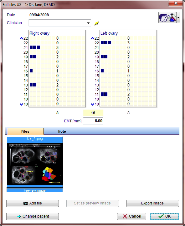
In the new window "Follicle US", you can enter the available follicles for the left and right ovary with millimetre accuracy.
| <img style="display: block; margin-left: auto; margin-right: auto;" src="/images/us1.png" alt="" width="581" height="714" /> |
|
The number of entered follicles per ovary will also be displayed numerically (left and right) and as total amount (middle). |
| <img src="/images/hint48.png" alt="" width="48" height="48" /> |
You can also add scanned photos or photos taken with a camera or other devices. |
The row labels stand for the following:
| Endom.[mm]: | SHH value |
| Total: |
Total number of follicles (left and right ovary) |
| LF left, LF right: |
Size of the leading follicles left and right [in mm]. |
| R-L above 22: | Number of follicles left/right with a diameter > 22mm. |
| Numbers 22-10: |
Follicles left/right with the diameter indicated by the number. |
| Below 10: |
Number of follicles left/right with a diameter < 10 mm. |
Deleting all ultrasound data
Right-click the ultrasound entry you wish to delete in the area “Follicle US” of the cycle overview. Click “Delete US” (the exact time is not relevant).
| <a href="/index.php?title=MedITEX_IVF_how_to">Back to How to</a> | <a href="#top">Back to top</a> |